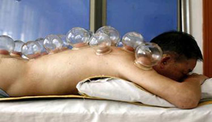
As a naturopath, nutritionist and healer, I was intrigued to explore the scientific reasons why the ancient art of cupping or Hijama therapy was so effective in treating a whole host of ailments and illnesses.
The wet cupping I found particularly fascinating and was curious to know more about the blood which was being extracted via the cup from various areas of the body.
From this viewpoint I started analyzing the blood under dark field and light field microscopy. This proved very insightful and proved to me something I had suspected.
The dry layered blood sample viewed under light field microscopy consistently showed high concentrations of toxic metals and chemicals, as well as showing evidence of bacterial and parasitic activity. The appearance of the live blood under the dark field microscope showed that there were high concentrations of acids and inflammatory proteins often referred to as fibrin.
These phenomena were more frequently present when the blood was removed from an area where the patient was experiencing pain and inflammation. I conclude from this that the area of pain appears to act like a magnet for acids, toxins and pathogens.
It is therefore very logical to assume that removal of these from the local area will bring about symptomatic relief, while encouraging fresh circulating blood to deliver healing nutrients and oxygen to the affected tissue, thus providing healing and resolution.
Together with dietary change, cleansing and detoxification therapy, along with education regarding the avoidance of toxins within the patient’s environment, I see Hijama or wet cupping, as a very effective adjunctive therapy on the path to wellness.
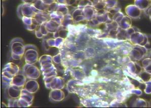
Proteinous waste found in live blood sample viewed under dark field microscopy. (This is often found when the diet contains too much cooked food and lacks enzymes for complete digestion).
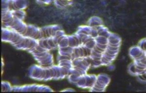
Rouleaux and Fibrin appearing in live blood under dark field microscopy. (This formation of red blood cells and inflammatory proteins are always present in blood that is overly acidic and infected).
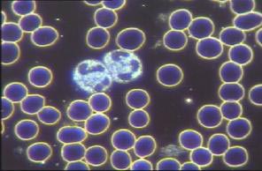
The ideal live blood picture under dark field microscopy Observe the red cells sitting separately in their own space, therefore having the freedom to travel around the body network of capillaries, which in some cases are one red cell in diameter. Observe the free-floating white blood cells known as neutrophils, again having the freedom to patrol around the body searching out toxins and pathogens.
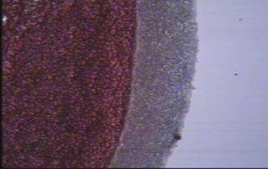
Toxic metals appearing on edge of dried blood sample under light field microscopy. (The thicker and denser the grey band, the more metals there are in the blood).
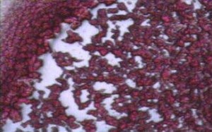
Degenerative processes evidenced under light field microscopy. (The white lakes seen here indicate that tissues associated with the vascular system have become inflamed and are harbouring atherosclerotic plaques, toxic metals and cholesterol).
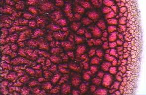
How the dry layered blood sample should look under light field microscopy (Observe the absence of any white lakes, the clean, finely cut edge showing no toxic metals and the black lines all joining together like a matrix).
Source: ICS Magazine BY David parker ND

